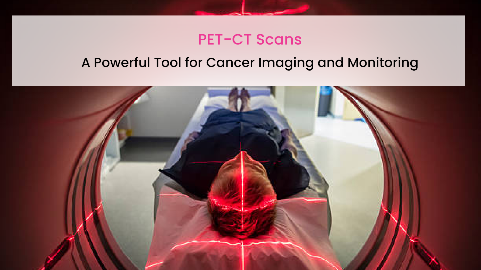
Positron emission tomography (PET) scans are a type of nuclear imaging test that can be used to diagnose, stage, and monitor cancer. PET scans work by using a radioactive tracer to create images of how tissues and organs are functioning. PET scans are often combined with computed tomography (CT) scans, which creates detailed images of the body’s anatomy. This combination of PET and CT scans, known as PET-CT, is a powerful tool for cancer imaging. PET-CT scans can provide more accurate information than either PET or CT scans alone, and they can help doctors to make better decisions about treatment.
Benefits of PET-CT Scans for Cancer Imaging and Monitoring
- Improved Sensitivity and Specificity: PET-CT scans have a higher sensitivity and specificity compared to standalone PET or CT scans. They can detect smaller tumours and metastases, enabling earlier detection of cancer and more accurate staging.
- Precise Anatomical Localization: PET-CT scans precisely localised areas of abnormal metabolic activity detected by PET to their exact anatomical location on the CT images. This is particularly useful in complex anatomical regions or areas where normal physiological activity can mimic cancer.
- Accurate Staging: Accurate staging is crucial for determining the extent of cancer spread. PET-CT scans allow better staging of cancer, helping clinicians make informed decisions about treatment options and prognosis.
- Assessing Treatment Response: PET-CT scans are essential for monitoring the effectiveness of cancer treatment. By comparing pre- and post-treatment scans, doctors can assess changes in tumour metabolism, size, and distribution, which aids in evaluating treatment response and modifying treatment plans if necessary.
- Early Treatment Evaluation: PET-CT scans can assess the effectiveness of cancer treatment earlier than traditional imaging methods. Early identification of treatment response allows for timely adjustments in treatment strategies.
- Whole-Body Imaging: PET-CT scans provide a comprehensive evaluation of the entire body in a single examination. This is particularly valuable for detecting distant metastases and assessing the overall disease burden in advanced cancer cases.Detecting Recurrence: PET-CT scans are highly sensitive in detecting cancer recurrence, even when it occurs at a small scale. Early identification of recurrent disease allows for prompt intervention and potentially better treatment outcomes.
- Research and Clinical Trials: PET-CT scans play a significant role in cancer research and clinical trials by providing reliable and quantifiable data on tumour characteristics, treatment response, and disease progression.
Tools for PET-CT scans:
- PET Scanner: The PET component of the scan uses a ring of detectors to capture the emission of positron-emitting radiotracers within the patient’s body. These scanners are sensitive to the gamma rays emitted during the decay of the radiotracer and can create a 3D image of the tracer distribution in the body.
- CT Scanner: The CT component of the scan utilises X-rays to provide detailed cross-sectional images of the patient’s anatomy. The CT images help in precise anatomical localization and fusion with the functional PET data.
- Radiotracers: Radiotracers are radioactive compounds that emit positrons (positively charged particles) during decay. These tracers are administered to the patient and accumulate in specific tissues or organs based on their metabolic activity. Commonly used radiotracers in PET-CT include FDG (Fluorodeoxyglucose) for general cancer imaging and various other tracers for specific cancers or conditions.
- Image Fusion Software: PET and CT images are acquired separately but need to be fused to create a single, comprehensive image. Specialised software is used to align and overlay the functional PET data onto the anatomical CT images, providing accurate localization of metabolic abnormalities within the patient’s anatomy.
- Picture Archiving and Communication System (PACS): PACS is a medical imaging technology that allows storage, retrieval, and sharing of the PET-CT images and reports. It is an essential tool for managing and accessing the vast amount of imaging data generated during PET-CT scans.
- Workstations and Viewing Software: Dedicated workstations equipped with specialised viewing software are used by radiologists and nuclear medicine physicians to analyse and interpret PET-CT images. These tools enable them to visualise the fused images, measure metabolic activity, and make diagnostic assessments.
- Radiation Safety Equipment: PET-CT involves the use of radioactive substances, and as such, radiation safety is a critical consideration. Protective gear, shielding, and monitoring equipment are used to ensure the safety of both patients and medical personnel.
PET-CT scans are a powerful tool for cancer imaging and monitoring. They offer a number of benefits over other imaging tests, including increased accuracy, improved staging, and better assessment of treatment response. PET-CT scans are generally safe and have few side effects.


